A Brief History of Protein Biosynthesis and Ribosome Research
by Hans-Jörg Rheinberger
Introduction
This topic cluster gives an overview of the history of protein synthesis, the structure and function of ribosomes, and of other major components of translation (see Refs. [1, 11-18] for earlier accounts). It refers to over twenty Nobel Laureates, but it also stresses and evidences the fact that many more researchers that also deserve to be remembered have contributed to that – ongoing – research endeavor. Not only a great number of scientists and research groups were involved in this work (see Refs. [2-10] for autobiographical accounts), but also widely different experimental systems, methods, and skills were used. Moreover, the efforts to elucidate the protein synthesis machinery were scattered all over the world. Nevertheless, a scientific community of surprising cohesion developed over time (cf. Refs. [19–26]). Emerging to a considerable degree out of cancer research at its beginning, the field of protein synthesis research gradually became an integral part of molecular genetics. To trace the broader context of the emergence of the experimental culture of translation research is the aim of this historically oriented topic cluster. My survey will mainly focus on the decades between the 1940s and the 1970s, the period that for most present-day readers constitutes something like a forgotten prehistory. The developments of the past decades will be treated more cursorily, since they are largely and excellently covered in the recent Lindau presentations of Ada Yonath, Thomas Steitz, and Venkatraman Ramakrishnan (Nobel Prizes for Chemistry 2009) (Ada Yonath, The Amazing Ribosome, 2010, Thomas Steitz, From the Structure of the Ribosome to (the Design of) New Antibiotics, 2011 and 2014, and Venkatraman Ramakrishnan, Seeing is Believing – A Hundred Years of Visualizing Molecules, 2015).
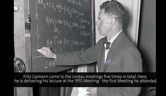
But first: a lesson in history. In May 1959, Paul Zamecnik, who must be regarded as the Nestor of protein synthesis research, was invited to deliver one of the prestigious Lectures at the Harvey Society in New York. He chose to speak about “Historical and current aspects of protein synthesis”, and he traced them back to “careful, patient studies” extending, as he said, “over half a century” [27, p. 256]. He then began with Franz Hofmeister [28] and Emil Fischer [29] (Nobel Prize for Chemistry 1902), who recognized the peptide bond structure of proteins; went on to Henry Borsook [30], who realized that peptide bond formation was of an endergonic nature; to Fritz Lipmann [31] (Nobel Prize for Physiology/Medicine 1953), who postulated the participation of a high-energy phosphate intermediate in protein synthesis; to Max Bergmann [32], who determined the specificity of proteolytic enzymes; to Rudolf Schoenheimer [33] and David Rittenberg [34], who pioneered the use of radioactive tracer techniques in following metabolic pathways; to Torbjörn Caspersson [35] and Jean Brachet [36], who became aware of the possible role of RNA in protein synthesis; to Frederick Sanger [37] (Nobel Prize for Chemistry 1958 and 1980), who unraveled the first primary structure of a protein, showing the specificity and uniqueness of the amino acid composition of insulin; and finally to George Palade [38] (Nobel Prize for Physiology/Medicine 1974), who gave visual evidence for the particulate structures in the cytoplasm acting as the cellular sites of protein synthesis. This is an impressive list of pioneers, who all, according to Zamecnik, “blazed the trail to the present scene” [27, p. 278]. The following offers a brief history of the quirks and breaks – continuing to the present - that marked protein synthesis research as an eminently collective and multidisciplinary endeavor.
The Archaeology of Protein Synthesis – The 1940s: Forgotten Paradigms
The early 1940s were the heydays of what Lily Kay [39] once aptly described as the ‘protein paradigm of life’. The transformation experiments of Oswald Avery and his colleagues at the Rockefeller Institute with DNA notwithstanding [40], proteins for quite some time continued to be seen as the key substances, not only of biochemical function, but also of hereditary transmission (from Delbrück [41] to Haurowitz [42]). It is surprising then to learn that, despite this early focus on proteins, the mechanism of protein synthesis largely remained a black box throughout the 1940s. Thoughts on mechanism during that decade mainly centered around the conception, favored by eminent biochemists of the time such as Max Bergmann and Joseph Fruton, that the mechanism of protein synthesis might be based on a reversal of proteolysis [43, 32, 44, 45]. The proteolysis concept, however, remained a controversial issue, especially since it could hardly be reconciled with the endergonic nature of peptide bond formation that appeared to be evident from Henry Borsook’s investigations at the California Institute of Technology in Pasadena. His measurements favored the idea that the formation of peptide bonds might involve some sort of activation of amino acids prior to their condensation, a topic on which Fritz Lipmann [31] as well as Herman Kalckar [46] had speculated as early as at the beginning of the 1940s. But despite the growing sophistication of experimental enzymology and of the structural, physical, and chemical analysis of proteins, including powerful new devices such as chromatography, electrophoresis, and X-ray crystallography (see Refs. [47, 48] for historical accounts), classical biochemistry alone and of itself did not provide a definite handle on the question of the cellular mechanisms of protein biosynthesis.
(00:00:16 - 00:05:00)
Some observations on the part of cytochemistry were intriguing but also remained erratic for the time being. Around 1940, Torbjörn Caspersson from Stockholm and Jack Schultz from the Kerckhoff Laboratories in Pasadena had developed techniques for measuring the UV absorption of nucleic acids within cells as well as UV microscopy of cells [35]. With that, they were able to correlate growth, i.e., the production of proteins, with the increased presence of ribonucleic acids at certain nuclear and cytoplasmic locations. Around the same time, Jean Brachet and his colleagues Raymond Jeener and Hubert Chantrenne in Brussels reached similar conclusions on the basis of differential staining and in situ RNase digestion of tissues [36].
The elucidation of the particulate structure of the cytoplasm by means of highspeed centrifugation dates back to the 1930s and derives from still other lines of research. Normand Hoerr and Robert Bensley in Chicago had used centrifugation to isolate and characterize mitochondria [49]. Albert Claude (Nobel Prize for Physiology/Medicine 1974), in James Murphy’s Laboratory at the Rockefeller Institute in New York, was working on the isolation of Peyton Rous’ chicken sarcoma agent when he, around 1938, incidentally realized that the particles he was sedimenting from infected cells had exactly the same chemical constitution than those sedimented from normal chick embryo tissue and thus were cellular constituents [50]. They were definitely smaller than mitochondria, and Claude termed them “microsomes” [51] accordingly. They were particularly rich in ribonucleic acid (Albert Claude, The Coming Age of the Cell, 1975, 17:30-26:00). Claude was tempted to place them in the category of ‘plasmagenes’ [52]. But they tenaciously resisted all attempts by Claude and his collaborators, especially Walter Schneider and George Hogeboom, to correlate specific and unique enzymatic functions with them [53, 54]. In contrast, however, the microsomes became preferential objects of ultracentrifugation. The centrifugation methods of Hubert Chantrenne [55] from Brachet’s laboratory in Brussels, and of Cyrus Barnum and Robert Huseby [56] from the Division of Cancer Biology at the University of Minnesota in Minneapolis pointed to a greatly varying size of the particles – if they had a definable size at all. But despite Brachet’s recurrent claim of a close connection between microsomes and protein synthesis, no particular experimental efforts were made in all these studies to enforce this line of argument [57]. However, the various efforts of an in vitro characterization of the cytoplasm by means of ultracentrifugation resulted in a set of procedures for the gentle isolation of cytoplasmic fractions [58] that soon proved very useful in a wide variety of other experimental contexts.
Basic Mechanisms – The 1950s
This situation was bound to change between 1945 and 1950 through still another approach to assess metabolic events. Right after World War II, radioactive tracers, especially 35S, 14C, and 3H as well as 32P became available for research to a wider scientific constituency as a byproduct of expanding reactor technology. The ensuing new attack on the mechanism of protein synthesis by way of radioactive amino acids benefitted greatly from the vast resources made available for cancer research after the War [59], and from the efforts of the American Atomic Energy Commission to demonstrate the potentials of a peaceful use of radioactivity [60, 61]. In fact, cancer research programs provided the background for much of the protein synthesis research during those years. This constellation also explains why much of protein synthesis research during the decade between 1950 and 1960 was done on the basis of experimental systems derived from higher animals, especially rat liver, and not on bacteria, as might be expected from hindsight.
Steps toward an in vitro Protein Synthesis System
The first attempts at approaching protein synthesis via tracing consisted in administering radioactive amino acids to test animals and in following the incorporation of the label into the proteins of different tissues. However, radioactively labeled amino acids were not yet commercially available at that time. One of the biggest concerns of these early radioactive in vivo studies was to maintain control over the specific activity of the injected material. Consequently, researchers in the field attempted to establish test tube protein synthesizing systems from animal tissues. Among the first to use tissue slices were Jacklyn Melchior and Harold Tarver [62], as well as Theodore Winnick, Felix Friedberg and David Greenberg [63], all from the University of California Medical School at Berkeley. Attempts to incorporate amino acids into proteins of tissue homogenates were also made at that time by Melchior and Tarver [62], by Friedberg et al. [64], and by Henry Borsook’s team at Caltech [65]. They all used different amino acids: sulfur-labeled cysteine and methionine (Tarver), carbon-labeled glycine (Greenberg and Winnick), and carbon-labeled lysine (Borsook). But some of the amino acid ‘incorporations’ in these early in vitro studies turned out to be due to amino acid turnover reactions. Granting that the experimental observation of amino acid ‘uptake’ indeed meant peptide bond formation became one of the biggest concerns of all those trying protein synthesis in the test tube between 1950 and 1955.
Paul Zamecnik’s work at the Massachusetts General Hospital (MGH) in Boston can rightly be considered to have been at the cutting edge of the field during the decade between 1950 and 1960. Zamecnik started his work on protein synthesis in 1945. In 1948, Robert Loftfield, an organic chemist from the Radioactivity Center at MIT, joined the staff of the Massachusetts General Hospital. In the preceding two years at MIT, he had worked out a suitable method for the synthesis of 14C-alanine and glycine [66]. Together with Loftfield, Warren Miller, and Ivan Frantz, Zamecnik started to introduce radioactive amino acids into slices of the livers of rats. In the laboratory of Lipmann, who was a neighbor of Zamecnik’s at MGH, William Loomis had just shown that dinitrophenol (DNP) specifically interfered with the process of phosphorylation [67]. When the Zamecnik group included DNP into one of their slice experiments, it stopped all amino acid incorporation activity. The result suggested that, as Lipmann had assumed for a long time on the basis of peptide bond model reactions, protein synthesis was indeed coupled with the utilization of phosphate bond energy [68].
(00:19:15 - 00:22:19)
There was no chance, however, to approach the problem by further manipulating liver slices. But to proceed along the lines of cell homogenization was a largely unexplored experimental field [69], and the MGH group worked for 3 years, from 1948 to 1951, to demonstrate the ‘incorporation’ via peptide bond formation of radioactive amino acids into protein in the test tube. In 1951, Philip Siekevitz, who had joined Zamecnik’s group in 1949, had achieved a preliminary fractionation of the liver homogenate by means of a regular Sorvall laboratory centrifuge [70]. None of the fractions was active when incubated alone. But when all of them were put together again, the activity of the homogenate was restored. In 1953, besides the introduction of a gentle homogenization procedure [71], the ordinary laboratory centrifuge was replaced by a high-speed ultracentrifuge. The new instrument made a quantitative sedimentation of the microsomes possible, leaving behind a non-particulate, soluble enzyme supernatant. Incorporation activity was restored from these two fractions under the condition that the test tube was supplemented with ATP and an ATP-regenerating system [72, 73]. It should, however, not be forgotten that the nucleus, too, was considered a site of protein synthesis throughout the 1950s; cf. e.g., Ref. [74]).
Amino Acid Activation and the Emergence of Soluble RNA
Toward the end of 1953, Mahlon Hoagland took up his work in Zamecnik’s lab, after having spent a year with Lipmann. Hoagland realized that he could use the technique of ‘phosphate-ATP-exchange’ developed in Lipmann’s lab (Fritz Lipmann, Protein Biosynthesis, 1967, 19:30-22:00) as a tool in Zamecnik’s rat liver system. A first partial, molecular model of protein synthesis resulted [75]. The combination of the phosphate exchange reaction with another model reaction, that of amino acids with hydroxylamine, suggested the activation by ATP of amino acids. What until then had simply been the ‘soluble fraction’, or the ‘105 000 x g supernatant’, or the ‘pH 5 precipitate’, became now viewed as a set of activating enzymes. Soon, other groups reported similar observations obtained in other systems. David Novelli, who had moved from Lipmann’s lab to the Department of Microbiology at Case Western Reserve University in Cleveland, established an amino acid-dependent PP/ATP-exchange reaction with microbial extracts [76]. Paul Berg (Nobel Prize for Chemistry 1980), from Washington University School of Medicine in St. Louis, reported on the activation of methionine in yeast extracts [77, 78]. Lipmann’s lab took up the task of isolating and purifying one of the amino acid-activating enzymes. Soon, the general character of the carboxyl-activation mechanism appeared to be established [79].
At this point, the participation of ribonucleic acids in protein synthesis still appeared as a black box, although microsomal RNA, by 1955, was generally assumed to play the role of an ordering device, jig, or ‘template’ for the assembly of the amino acids. The actual point of discussion at that time, however, to which Sol Spiegelman from the University of Illinois at Urbana and Ernest Gale from Cambridge repeatedly referred, was accumulating evidence for a coupling of the synthesis of proteins with the actual synthesis of RNA [80–82]. That same year, Marianne Grunberg-Manago in Severo Ochoa’s (Nobel Prize for Physiology/Medicine 1959) laboratory in New York identified an enzyme which was able to synthesize RNA from nucleoside diphosphates [83].
Late in 1955, Zamecnik, too, began to look for RNA synthesis activity in his fractionated protein synthesis system. He added radioactive ATP to a mixture of the enzyme supernatant and the microsomal fraction. In a parallel experiment, Zamecnik had incubated non-radioactive ATP and 14C-labeled leucine instead of non-radioactive leucine and 14C-labeled ATP together with the fractions. As Zamecnik recorded in his notebook, the assay suggested that radioactive leucine as well became attached to the RNA [84]. Zamecnik had found an RNA in the soluble fraction to which amino acids were attached. The new entity was termed ‘soluble RNA’.
(00:21:56 - 00:26:01)
The further differentiation of the cell-free protein synthesis system now became the working field for a growing protein synthesis ‘industry’. In 1956, Robert Holley (Nobel Prize for Physiology/Medicine 1968), from Cornell University, gathered evidence for the presence of an RNA intermediate in protein synthesis [85]. Paul Berg, soon joined by James Ofengand, went ahead with studies on the amino acid incorporation into soluble RNA of Escherichia coli [86]. Tore Hultin from the Wenner-Gren Institute in Stockholm obtained independent evidence for an intermediate step in protein synthesis from kinetic isotope dilution studies [87]. Kikuo Ogata and Hiroyoshi Nohara at the Niigata University School of Medicine in Japan also had collected hints for an RNA-connected intermediate in protein synthesis [88]. By the end of 1957, amino acid–oligonucleotide compounds were being investigated by at least three other research groups: Victor Koningsberger, Olav Van der Grinten, and Johannes Overbeek [89] at the Van’t Hoff Laboratory in Utrecht; Richard Schweet, Freeman Bovard, Esther Allen, and Edward Glassman [90] at the Biological Division of Caltech; and Samuel Weiss, George Acs, and Fritz Lipmann [91], who had moved from the Massachusetts General Hospital to the Rockefeller Institute in New York. All of them joined the race for adding items to the list of what these molecules and their activating enzymes did and what they failed to do. What had emerged as a biochemical intermediate in protein synthesis soon turned into one of those big missing pieces within the flow scheme of the expression of molecular information. At Richard Schweet’s suggestion, the molecule was later referred to as transfer RNA [92], and it became identified with what, based on considerations rooted in the double-helical structure of DNA, Francis Crick (Nobel Prize for Physiology/Medicine 1962) had postulated as an adaptor of the genetic code [93–95].
From Microsomes to Ribosomes
As we have seen, it was not until the beginning of the 1950s, and in a context quite different from their original characterization, that the ‘small particles’ or ‘microsomes’ became linked to protein synthesis in vivo [97–102] and in vitro [70, 96, 102, 103]. But it took another decade before the isolation of active cytoplasmic particles through sucrose-gradient centrifugtion became a laboratory standard. To obtain ‘purified’ microsomes became one of the major issues in the development of cell-free protein synthesis [104]. For purification, Zamecnik’s colleague John Littlefield took advantage of the detergent sodium deoxycholate that solubilized the protein–lipid aggregates of the microsomal fraction. The RNA-to-protein content (1 : 1) of his particles corresponded to those of Howard Schachman from Wendell Stanley’s (Nobel Prize for Chemistry 1946) Virus Laboratory in Berkeley for Pseudomonas fluorescens [105] and by Mary Petermann from the Sloan Kettering Institute in New York for rat liver and spleen [106].
Around the same time, George Palade [38] was able to visualize small, electron dense particles on the surface of the endoplasmatic reticulum in situ by means of electron microscopy. Siekevitz had joined Palade in 1954. He added his biochemical expertise to the work at the Rockefeller Institute [107]. Besides electron microscopy, the calibration of these ‘macromolecules’ involved velocity sedimentation and electrophoretic mobility [106, 108–110]. They became a synonym for cytoplasmic RNA, although the postmicrosomal supernatant invariably also contained RNA – approximately 10% of the cell’s total RNA [107]. From analytical ultracentrifugation, a sedimentation coefficient of the particles could be calculated.
In the course of the 1950s, the RNA of these particles was generally assumed to provide the template upon which the amino acids were assembled into proteins. In 1958, Howard Dintzis coined the term ‘ribosome’ for purified microsomes devoid of membrane fragments (Wim Möller, pers. comm.; see also Refs. [111, 112]). During the following years, this neologism made its way into the laboratories and into the literature. The reason for changing the name was the presumed role of the particle’s RNA. The new designation no longer reflected a mere technical representation, but a biological function. Like ‘transfer RNA’, the ‘ribosome’ began to relocate protein synthesis from biochemistry to molecular genetics, transforming it into an integral part of what Crick, apparently without minding about the theological connotations of the term, had called the “central dogma” of molecular biology [113, p. 153]. It codified the notion that the genetic information makes its way from DNA to RNA to protein. The central dogma subsumed the process of protein synthesis as the final, translational, step in the overarching process of gene expression.
With respect to their physical parameters, the protein synthesizing particles considerably changed their appearance between 1955 and 1960. Around 1956 and after many trials, Schachman had found yeast microsomes sedimenting with a velocity constant (S) of 80 and to dissociate reproducibly into two unequal portions of 60S and 40S [114]. In a similar manner, Petermann and co-workers were able to separate 78S liver ribosomes into 62S and 46S particles [115]. Alfred Tissières and James Watson (Nobel Prize for Physiology/Medicine 1965), at Harvard, had their bacterial E. coli particles sediment with 70S. They could be dissociated reversibly into a 50S and a 30S component [116, 117]. It was realized that the secret of stabilization lay chiefly in the concentration of divalent Mg2+ ions. Work on a variety of particles from other sources began to converge on two distinguishing features: bacterial particles (roughly 70S) were consistently smaller than their eukaryotic counterparts (roughly 80S), but both could be separated into something that began to be recognized as a small and a large ribosomal subunit.
Models
The state-of-the-art of protein synthesis, as a process of translation of genetic information, was conceptually re-framed by Francis Crick and his colleagues, especially Sydney Brenner (Nobel Prize for Physiology/Medicine 2002), between 1955 and 1957, and summarized by Crick in his seminal paper of 1958. After years of theorizing from template models, starting with Hans Friedrich-Freksa [118] and Max Delbrück [41] (Nobel Prize for Physiology/Medicine 1969), and continuing with Hubert Chantrenne [119], Felix Haurowitz [42], Alexander Dounce [120], Victor Koningsberger and Johannes Overbeek [121], Fritz Lipmann [122], George Gamow [123], Henry Borsook [124] and Robert Loftfield [125], Crick had come up with a new proposal. During the 1940s, models of autocatalytic protein replication had been at the forefront. Gene duplication thus meant protein duplication. Later models conceived the process of molecular information transfer in terms of a physicochemical interaction between ribonucleic acids and amino acids involving covalent bonding. In the aftermath of the Watson and Crick [126] seminal model of the DNA double helix, Gamow [123] proposed an interaction between DNA and amino acids based on the geometrical shape of holes in the double helix. Crick, thinking of the complementarity features of the DNA double helix, now envisaged what he called “adaptation”, i.e., a specific base-pairing interaction between an amino acid-carrying nucleic acid adaptor exposing a signature complementary to the code of a template nucleic acid. It is interesting to note that at the time Crick launched his adaptor hypothesis, he obviously did not judge it important enough to be published. It was only its linkage to soluble RNA that made it a prominent concept and a prophecy as seen in hindsight.
With the surprising emergence of soluble, amino acid-carrying RNAs, a new possibility of cracking the code seemed to appear on the horizon. Meanwhile, Zamecnik and Liza Hecht had established as a common feature of all S-RNAs a common 3’-end to which the amino acids became attached: an invariable -CCA trinucleotide [127]. This was anything but a distinct code! Hoagland had hoped to have, with transfer RNA, the “Rosetta Stone” for deciphering the code in his hands [128, p. 61]. But trying to obtain the code through transfer RNA with a direct experimental approach led only to a dead end. Ernest Gale and Joan Folkes at Cambridge, who were analyzing the relation between protein synthesis and the synthesis of nucleic acids in a staphylococcal in vitro system, also got stuck [129–131]. Robert Holley, who since 1957 had put all his efforts into isolating, purifying and sequencing the SRNA specific for alanine from yeast, needed many years and a massive crew of coworkers to arrive at the primary sequence of the first transfer RNA [132]. When he presented the sequence, the code had been solved on a completely different experimental track.
The Golden Age of Translation – The 1960s
The genetic code was solved between 1961 and 1965 with a breathtaking velocity that nobody would have dared to predict. The 1960s also saw the emergence of messenger RNA, the dissection of the ribosome into its components, and the resolution of the translational process into partial functions. Through transfer RNA, messenger RNA, and the code, the biochemistry of protein synthesis merged and for a while even tended to become synonymous with molecular biology, a situation that had been unimaginable a decade earlier when a gap still loomed large between those who considered themselves to be the avantgarde of molecular biology and those who did the messy work of experimentally draining the ‘bog’ of nucleic acid or protein biochemistry and metabolism [10]. In vitro systems remained central to the field. Of particular importance was the transition from mammalian systems to bacterial, especially E. coli systems of protein synthesis. E. coli, so vital as a genetic model throughout the 1950s, was not yet a model of translation during this decade. It was only around 1960 that molecular genetics and protein synthesis research joined forces on the basis of one single model organism (see Ref. [133] for an overview of the field around 1960).
From Enzymatic Adaptation to Gene Regulation:
Messenger RNA
Toward the end of the 1950s, the work of Jacques Monod (Nobel Prize for Physiology/Medicine 1965) and François Jacob (Nobel Prize for Physiology/Medicine 1965) at the Pasteur Institute in Paris resulted in a major contribution to understanding protein synthesis and its regulation. Since the beginning of the 1940s, Monod had studied ‘enzyme adaptation’ in E. coli [134]. Around 1955, he and his co-worker Georges Cohen distinguished three genes: the y-gene specifying a permease responsible for the import of lactose into the bacterial cell, the z-gene responsible for the sugar-decomposing ß-galactosidase, and an i-factor responsible for the induction of the system. François Jacob had started his work on the viral phenomenon of lysogeny in the laboratory of André Lwoff (Nobel Prize for Physiology/Medicine 1965) at the Pasteur Institute in 1950. Around that time, decisive developments in bacterial genetics were about to take shape. William Hayes in London and Luigi Cavalli-Sforza in Milan found hints for a sexual differentiation in E. coli bacteria and learned to distinguish between donor and recipient cells during conjugation. In 1953, Hayes in addition characterized a high-frequency recombinant donor variant of E. coli (K12). In the process of doing recombination kinetics with multiple mutants of K12, Jacob and Eli Wollman invented a trick that proved to be highly consequential: If the process of conjugation was interrupted at certain time intervals by mechanical agitation in a mixer, the transfer of different characters could be resolved in a linear fashion. ‘Mapping by mating’ became a clue to the genetic mapping of bacterial chromosomes [135].
In 1957, Monod and Jacob joined forces and included Arthur Pardee from the virus laboratory of the University of California at Berkeley in their efforts. They resulted in the famous series of experiments that led to the operon model of gene expression. The experiments suggested the involvement of a cytoplasmic repressor and a functionally “unstable intermediate” responsible for the expression of the structural genes [136, p. 224]. When Jacob and Sydney Brenner drew a parallel between these experiments and the observation of Lazarus Astrachan and Elliot Volkin [137] from the Oak Ridge National Laboratory of a quickly metabolizing RNA that appeared after infection of their bacteria with T2 phages, the question became whether this unstable intermediate was some sort of an “information carrying RNA” [136, p. 225] that transiently combined with existing microsomes, thus inducing the immediate synthesis of a specific protein [6]. There had been hints in the literature pointing towards quickly metabolizing RNAs for quite some time. As early as 1955, microbiologist Ernest Gale in Cambridge had claimed that in inducible systems, protein synthesis is accompanied by or even dependent upon RNA synthesis [138]. In addition, Sol Spiegelman, who also worked on enzyme induction, had assumed that the RNA templates of induced enzymes are unstable [139].
The concept of microsomes had emerged from eukaryotic in vitro systems with reduced metabolic activity, and as it had gained currency towards the end of the 1950s, it was clearly at odds with these observations on bacterial metabolism. Microsomal RNA appeared to be inert, and for all those working on cells from higher organisms, the ribosome represented “a stable factory”, already containing an RNA transcript of DNA [10, p. 107]. Moreover, bacterial in vitro systems had a bad reputation in the leading circles of protein synthesis workers in the late 1950s. They were considered ‘dirty’ systems that were difficult to control [125].
The decisive experiment establishing the role of messenger RNA came from a joint effort of Jacob, Brenner and Matthew Meselson at Caltech: They grew bacteria on heavy isotopes to tag the ribosomes and infected the E. coli cells with a virulent phage in the presence of radioactive isotopes. What they found was that newly synthesized radioactive phage RNA indeed became associated with pre-existing heavy ribosomes [140]. ‘Messenger RNA’ [141] now assumed the general meaning of a molecular information transmitter whose transcription was controlled by feedback loops according to the operon model. Around the same time, Masayasu Nomura and Benjamin Hall, in Spiegelman’s laboratory at Urbana, had characterized a ‘soluble’ form of RNA synthesized in E. coli after bacteriophage T2 infection. It became associated with ribosomes in the presence of high magnesium concentrations [142]. And François Gros, Walter Gilbert (Nobel Prize for Chemistry 1980), and Chuck Kurland, in the laboratory of Watson at Harvard, showed that unstable ‘messenger RNA templates’ also belonged to the metabolic makeup of uninfected E. coli cells [143].
A Bacterial in vitro System of Protein Synthesis and the
Cracking of the Genetic Code
The differentiation of reliable bacterial in vitro systems occurred in parallel, but independent of the experimental context of enzyme induction. The first to report on a system based on E. coli were Dietrich Schachtschabel and Wolfram Zillig at the Max Planck Institute for Biochemistry in Munich [144]. In 1958, Marvin Lamborg, a postdoctoral Fellow of the National Cancer Institute from NIH, had come to work with Zamecnik. Lamborg finally managed to establish a cell-free protein synthesis system based on E. coli extracts [145]. In a rapid dissemination, the Lamborg–Zamecnik type of system made its way into other laboratories and soon became a model system for protein synthesis research. Besides Tissières in Watson’s lab [146], among the first to use such a system were David Novelli at the Oak Ridge National Laboratory [147], Daniel Nathans and Fritz Lipmann [148] at the Rockefeller Institute in New York, Kenichi Matsubara and Itaru Watanabe [149] at the University of Tokyo and at Kyoto University, and James Ofengand, then on a fellowship at the Medical Research Council Unit for Molecular Biology in Cambridge [150]. In Watson’s group, the structure and function of bacterial ribosomes and messenger RNA had moved to the center of attention. But the E. coli system was also being introduced at the National Institutes of Health in Bethesda. The days of the rat-liver system as a pace-maker for unprecedented events were over.
Marshall Nirenberg (Nobel Prize for Physiology/Medicine 1968), at NIH, had just started to establish a cell-free E. coli system when Heinrich Matthaei joined him in the fall of 1960. Nirenberg had set himself the task of investigating the steps that connect DNA, RNA and proteins, and synthesizing, in a cell-free system, a specific protein [151]. Despite many efforts (cf., e.g., Ref. [152]), the synthesis of a defined and complete protein in vitro had remained a challenge ever since the end of the 1940s. To begin with, Nirenberg and Matthaei proceeded well within the context of the prevailing picture of the ribosome, its RNA still being assumed to play the role of a template. A minor, but finally decisive procedure set the stage for their accomplishment: the preincubation of the bacterial cell extract. Matthaei and Nirenberg put the system to work until its endogenous activity came to a halt. Then, according to a principle of variation, they introduced additional RNAs into the system. It was a lucky coincidence that the synthesis of RNA fell into the special expertise of Leon Heppel, who was the director of the laboratory in which Nirenberg and Matthaei were working. With these polymers at their disposal, they needed only a few months until they, by systematically varying their radioactive amino acids, had deciphered the first code word: The homopolymer polyuridylic acid coded for the artificial protein poly-phenylalanine [18, 153].
We have thus to assume that the concept of messenger arose at least twice in the history of molecular biology. It emerged from two experimental contexts that could not have been more different: from a delicate, genetically triggered in vivo system of enzyme induction, and from a comparatively modest, fractionated in vitro system of protein synthesis. Despite these radical breakthroughs, microsomal RNA continued to be considered for quite a while as a possible template [154].
After the Fifth International Congress of Biochemistry in August 1961 in Moscow, where Nirenberg reported the findings from his laboratory, the other attempts at deciphering the code by genetic and chemical microanalysis of phage mutants in Cambridge and of tobacco mosaic virus mutants in Berkeley and Tübingen could be dropped (see, e.g., Refs. [155–158]). The subsequent hunt for the different code words became a matter of refining the experimental conditions of the E. coli system. The triplet-binding assay of Philip Leder was one of the key accomplishments in the years to come [159]. Besides Nirenberg, it was mainly Severo Ochoa and his co-workers in New York and Gobind Khorana (Nobel Prize for Physiology/Medicine 1968) in Wisconsin who, on the basis of their experience with polymer synthesis, were able to join the race ([160], see Ref. [161] for a review). An initiation codon and the corresponding, formylated initiator tRNA [162, 163] as well as special codons functioning as stop signals were soon identified genetically [164, 165] and biochemically [166–168]. By 1967, the complete code was in place. For the next 10 years, the new findings on translation resulted, along the lines of ever-new twists, quirks, and refinements, from the dissection of bacterial systems [19].
The Functional Dissection of Translation
With the isolation of ribosomes, the purification of specific transfer RNAs and their corresponding synthetases, and the beginning of a deliberate manipulation of viral and synthetic messengers, the stage was set for the dissection of ribosomal function [169, 170]. From the first observations onwards [171], one of the big riddles concerning the energy turnover of peptide elongation had been the involvement of GTP in the process. Around 1960, it had become clear that GTP was not involved in the amino acid-activation reaction per se. In a manner still not understood, GTP did interfere with the amino acid transfer mechanism (see discussion in Ref. [172]). The transfer depended on a partial fraction of the pH 5 enzyme supernatant [173]. It took another three years until Jorge Allende and Robin Monro in Lipmann’s lab identified an enzyme fraction in E. coli whose transfer activity overlapped with a GTPase activity [174] and was termed ‘G factor’ ([175, 176], see Ref. [3] for a review). At the same time, a complementary ‘T factor’ was resolved into a temperature-stable component Ts and an unstable component Tu [177].
(00:41:58 - 00:50:03)
In bacteria, they became known as elongation factors EF-G and EF-Tu/EF-Ts (EF2 and EF1A/EF1B, respectively, in eukaryotes). In a reticulocyte system, Boyd Hardesty and Richard Schweet, a few years earlier, had already identified two fractions, TF-1 and TF-2, that were involved in the GTP-dependent interaction of Phe-tRNA with poly(U)-programmed ribosomes [178]. The identification of three factors required for the initiation [179, 180], and of factors required for the termination of the translation process soon followed [181].
Transfer RNA binding to ribosomes and to their subunits became a major subfield for studying ribosomal function. Among the pioneers were Tore Hultin in Stockholm and Leendert Bosch in Leiden [182–185]. Around the same time, Hoagland found that uncharged S-RNA bound to the microsomes as well as did S-RNA charged with amino acids [186]. The majority of the ensuing tRNA binding studies was done in bacterial systems, where the poly(U)-dependent Phe-tRNA binding assay became by far the most prominent. Soon, Walter Gilbert showed that the tRNA carrying the growing polypeptide is associated with the 50S subunit [187], whereas the binding of poly(U) apparently involved the small subunit [188], and the binding of transfer RNA in general depended on the presence of a messenger [189]. Jonathan Warner and Alex Rich found active reticulocyte ribosomes carrying two transfer RNAs [190].
A functional and clearcut distinction between two different binding sites of charged tRNAs on the ribosome was still missing. Robert Traut and Robin Monro [191] provided it with the puromycin-peptidyltransferase assay which allowed investigators to distinguish a puromycin-sensitive (P-site) and a puromycin-insensitive binding state (A-site) of aminoacylated tRNA. Based on this observation, the two-site model of ribosomal elongation became codified by Watson [192] and continued to serve as a reference system for research on ribosomal function well into the 1980s. Many features of translational initiation [193], elongation [194–196], and termination [197] were outlined in more and more sophisticated and reduced partial in vitro systems [198].
(00:32:57 - 00:36:05)
(00:30:51 - 00:37:53)
Antibiotics revealed themselves to be invaluable tools for the dissection of partial ribosomal functions as well as for the ongoing in vivo studies concerning regulation, speed, and accuracy of protein synthesis. Among the prominent drugs were puromycin as an elongation terminating agent (see Refs. [199–201] for early studies); chloramphenicol as a specific inhibitor of bacterial peptidyltransferase [202–204]; fusidic acid as interfering with the translocation factor EF-G [205, 206]; and streptomycin as inducing misreading [207, 208]. One of the earliest realistic measurements concerning the accuracy of the process of polypeptide formation came from Robert Loftfield [209].
(00:24:52 - 00:32:23)
(00:16:56 - 00:27:54)
In the context of pursuing ribosomal function, and after the mRNA concept had been established, gentle isolation of messenger–ribosome complexes became a matter of priority in the early 1960s. Particles larger than 70S or 80S appeared on sucrose-gradient patterns and electron microscopic images. For them, the term ‘polysomes’ quickly came into general use [210-214]. They appeared to consist of strings of ribosomes occupying a particular messenger RNA. Special isolation procedures were required to prevent them from breaking down to monosomes during fractionation. On the other hand, in vivo and in vitro evidence grew that ribosomes dissociated and reassociated during their functional cycle [215, 216], and that initiation started on the 30S subunit [217].
Around the same time, Peter Traub, together with Nomura, found the right temperature and ionic conditions for reconstituting the small ribosomal subunit of E. coli in the test tube [218] from its RNA and protein moieties, respectively. The 50S subunit assembly proved more difficult [219] but was finally achieved by Knud Nierhaus and Ferdinand Dohme [220]. After the much simpler, symmetric TMV in the early 1950s, the two highly asymmetric ribosomal subunits became the emblem of molecular self-assembly in the late 1960s and early 1970s [219, 220]. The possibility of in vitro ribosome assembly opened the field for a multiplicity of structure–function correlation studies at a previously unknown level. The hope, however, that a particular ribosomal protein might be singled out as responsible for the peptidyl transferase reaction did not materialize.
The Structural Dissection of the Ribosome
The original assumption of Watson at Harvard, Schachman in Berkeley, and others who started to analyze the structure of bacterial particles, had been that it might be analogous to that of RNA viruses: an RNA moiety wrapped with multiple copies of a coat protein. The analogy had certainly not been favorable either to the emergence of the concept of messenger RNA, or to the emergence of the view of an asymmetric particle consisting of many different proteins.
In view of the complex protein make-up of ribosomes, it is not surprising that their RNA moiety was the first component to be characterized in terms of sedimentation behavior, molecular weight, and overall base composition. As for the ribosomes, so for rRNA, too, sucrose-gradient centrifugation was crucial. Around 1960, there was still considerable uncertainty about the identity of ribosomal RNA. Before RNase-free strains of bacteria became available [221, 222], the problem of RNA breakdown during preparation could hardly be mastered. The introduction of the separation of RNA from cellular protein by phenol extraction greatly facilitated laboratory manipulation of RNA. This method came into quick and general use soon after its publication [223, 224]. In 1959, Paul Ts’o [225] separated rRNA from pea seedlings and rabbit reticulocytes into two major 28S and 18S peaks. A series of careful studies on E. coli ribosomes in Watson’s laboratory led Kurland to propose that ribosomal RNA came in two large species, 16S and 23S, respectively [226]. Alexander Spirin in Moscow had reached basically the same conclusion [227]. The question however whether this represented the ‘native’ state of ribosomal RNA, whether originally they were made up from smaller fragments or derived from a large precursor, continued to be a matter of debate for several years [228]. The controversy eventually came to a satisfactory end when it became evident that mature ribosomal RNA originated from a large transcript that was processed in the event of ribosome formation, and that, indeed, a small defined RNA, 5S RNA, was part of the 50S subunit [229]. Subsequently, 5S rRNA became the first ribosomal RNA molecule to be completely sequenced in 1968 [230]. This breakthrough had been made possible through Sanger’s 2D fractionation procedure for radioactive nucleotides [231]. It took three years to determine its 120 nucleotides. In comparison, sequencing the first transfer RNA (yeast tRNAAla) with slightly more than half the number of nucleotides had taken Holley and his co-workers some eight years [132]. Other groups soon followed with other tRNA species [232, 233]. The detailed functional elucidation of these molecules, however, had to await further studies. Their crystallization proved to be a major prerequisite for moving forward in this direction (see Refs. [234, 235] among others).
Serious analysis, on the basis of starch-gel electrophoresis, of the protein composition of ribosomes goes back to the work of Jean-Pierre Waller [236] and to the fractionation studies of David Elson [237] and Pnina Spitnik-Elson [238]. One of the first ribosomal proteins to be characterized individually was the acidic A-protein of the large subunit studied by Wim Moeller and later known as L7 (L12) [239]. Major efforts to develop methods for separating and purifying individual proteins came, among others, from Heinz Günter Wittmann and Brigitte Wittmann-Liebold’s laboratory in Berlin [240], Tissière’s in Geneva [241], and Kurland’s in Wisconsin [242]. A prominent achievement in this endeavor was the separation of all ribosomal proteins by 2D polyacrylamide gel electrophoresis [243]. It served as an efficient and economizing standardization vehicle in the field of ribosomal protein identification.
1970s–1990s: A Brief Synopsis
The survey of the following three decades from the 1970s to the 1990s will be very brief. There is no need to go into the details of an ongoing research in this historical introduction, since the major events during these decades are extensively dealt with in some recent Lindau lectures. The 1970s can be regarded as the period of the elucidation of the primary structure of the components of the translational apparatus. Indeed, around the turn of the decade, the ribosome of E. coli became the first cellular organelle whose RNA [244–246] and protein components [247] were completely sequenced. Sequencing the complete ribosomal RNA became a feasible task only after the new sequencing methods of Maxam and Gilbert [248], and of Sanger [249] had been introduced.
The emergent recombinant DNA technologies helped to construct a detailed genetic map of the components involved in protein biosynthesis. The ribosomal RNA genes, however, were mapped before the era of recombinant DNA technology. A dozen years had elapsed between their first identification in 1962 [250] and their precise mapping [251]. Knowledge about ribosomal protein genes and operons rapidly accumulated after the subsequent isolation of protein gene-transducing lambda phages [252]. Another source of information was provided by the systematic work with ribosomal protein mutants [253, 254].
Molecular details of ribosomal function also became available, such as the interaction of mRNA with 16S RNA during initiation [255, 256], and the mechanisms by which ribosomes achieve their accuracy [257, 258]. The regulation of ribosome biosynthesis, starting with the early findings on the genetics of RNA synthesis [259], also became a major field of investigation during the 1970s [260–262]. Over the years, a detailed view, first of transcriptional, then of translational feedback regulation mechanisms emerged. Since Monod and Jacob’s work on the lac operon, transcriptional control had been the leading paradigm. The shift of interest from transcriptional to translational regulation was indeed an unprecedented turn, both in prokaryotes and in eukaryotes (for the latter, see Severo Ochoa, The Regulation of Protein Synthesis in Reticulocytes, 1978). The major events in this area have both been initiated and reviewed some time ago by Nomura [8].
During the 1970s, ribosome research became a focus for the development and application of numerous advanced biochemical, biophysical and biological techniques. In vitro reconstitution of ribosomes [263] and in situ localization of ribosomal components via immunoelectron microscopy [264, 265], scattering studies [266], cross-linking [267] and affinity labeling [268] led to early insights into the quaternary structure of the protein synthesizing organelle and its functional characteristics such as factor binding and the constitution of the peptidyltransferase center.
The 1980s, on the one hand, were characterized by an increasing backshift of emphasis towards eukaryotic systems (see Ref. [269] for a contemporary overview). Ira Wool in Chicago had pioneered mammalian ribosomal proteins during the era of E. coli (see Ref. [270] for a review), Rudi Planta in Amsterdan had done much of the genetic and structural work on yeast ribosomal RNA (see Ref. [271] for a review). On the other hand, after a lag period, the tedious and time-consuming task of secondary, tertiary, and quaternary structure modelling came to fruition and became linked to ribosomal function. Protein–protein crosslinking [272], protein–RNA crosslinking [273, 274], protection and modification studies [275], neutron scattering [276, 277], electron microscopy [278], and ribosome crystallization [279] figure prominently among the methods involved in this continuing endeavor. On the functional side, exhaustive tRNA-binding studies led to new model conceptions of the elongation cycle involving a third tRNA binding site [280–283, 275]. Peter Moore has judged on this topic: “The two-site model for the ribosome, which the world has accepted for a generation is dead. The existence of a third site for tRNA binding, the exit site, is now established beyond reasonable doubt. This is unquestionably the most significant advance in our understanding of the ribosomal events of protein synthesis in many years” [284].
Finally, the 1990s were dominated by major efforts to carry the structural analysis of ribosomes to atomic resolution. The availability of suitable crystals of ribosomes and ribosomal subunits, particularly from thermophilic and halophilic sources, and the solution of the phasing problem led to a proliferation of X-ray crystallographic studies to which Wittmann and Ada Yonath [285] in Berlin and the group at Pushchino [286] had laid the ground with their ribosome crystallization initiatives in the 1980s. After almost 20 years of continued efforts, atomic resolution has now been achieved for the large ribosomal subunit from Haloarcula marismortui [287] and Deinococcus radiodurans [288], the small subunit from Thermus thermophilus [289, 290], and near-atomic resolution for the 70S-tRNA–mRNA complex [291]. In parallel, the development of cryoelectron microscopic image reconstruction has helped to refine the overall 3D shape of ribosomal particles, in particular as related to specific functional states [292, 293]. Thus a dynamic picture of the ribosome at atomic resolution emerged around 2000 for which the Nober Prize in Chemistry was awarded to Ada Yonath, Thomas Steitz, and Venkatraman Ramakrishnan in 2009.
(00:07:28 - 00:31:00)
(00:04:59 - 00:08:48)
Evidence of the involvement of rRNA in ribosomal functions has become overwhelming over the past two decades. Seminal in this context was certainly Carl Woese with his speculations on the origin of the protein synthetic machinery [294]. But it was the characterization of catalytic activities of precursor ribosomal RNA initiated by Thomas Cech (Nobel Prize for Chemistry 1989) [295] that turned the ‘protein paradigm’ of the ribosome, prevalent in the 1960s and 1970s, back into an ‘RNA paradigm’. (Indeed, in the early days of ribosomology, rRNA had been closely associated with ribosomal function. That function — of a template — however, did not survive history.) Indications accumulated that 23S RNA is involved in the peptidyltransferase reaction [296, 297], which until then was thought to be a domain of the ribosomal proteins. Efforts to achieve peptidyltransfer activity with ribosomal RNA alone have so far not been successful [298, 299]. The atomic model of the 50S subunit now appears to suggest that ribosomal RNA may indeed be able to do the job without direct involvement of proteins [300]. On this view, the ribosome finally turned out to be a veritable ribozyme, and there is now evidence at the atomic level concerning the nature of the catalytic mechanism of the peptidyltransfer reaction.
(00:15:30 - 00:23:39)
(00:15:30 - 00:23:39)
References
1 H.-J. Rheinberger: Toward a History of Epistemic Things. Synthesizing Proteins in the Test Tube, Stanford University Press, Stanford 1997.
2 P. C. Zamecnik, An historical account of protein synthesis, with current overtones – a personalized view,
Symp. Quant. Biol. 1969, 34, 1–16.
3 F. Lipmann: Wanderings of a Biochemist, Wiley-Interscience, New York 1971.
4 P. C. Zamecnik, Historical aspects of protein synthesis, Ann. NY Acad. Sci. 1979, 325, 269–301.
5 P. Siekevitz, P. C. Zamecnik, The ribosome and protein synthesis, J. Cell. Biol. 1981, 91 (3), 53S–65S.
6 F. Jacob: The Statue Within, Basic Books, New York 1988.
7 F. H. C. Crick: What Mad Pursuit, Basic Books, New York 1988.
8 M. Nomura: History of Ribosome Research: A Personal Account, in The Ribosome. Structure, Function and Evolution, eds W. E. Hill, P.B. Moore, A. Dahlberg et al., ASM Press, Washington, DC 1990, 3–55.
9 A. Spirin: Ribosome Preparation and Cell-free Protein Synthesis, in The Ribosome. Structure, Function andEvolution, eds W. E. Hill, P.B. Moore, A. Dahlberg et al., ASM Press, Washington, DC 1990, 56–70.
10 M. Hoagland: Toward the Habit of Truth, W. W. Norton and Company, New York 1990.
11 F. H. Portugal, J. S. Cohen: A Century of DNA, The MIT Press, Cambridge, MA 1977.
12 H. F. Judson: The Eighth Day of Creation, Simon and Schuster, New York 1979.
13 H.-J. Rheinberger: Experiment, difference, and writing. I. Tracing protein synthesis, and II. The laboratory production of transfer RNA, Stud. Hist. Philos. Sci. 1992, 23, 305–331, 389–422.
14 H.-J. Rheinberger, Experiment and orientation: early systems of in vitro protein synthesis, J. Hist. Biol. 1993, 26, 443–471.
15 H.-J. Rheinberger, From microsomes to ribosomes: ‘strategies’ of ‘representation’, J. Hist. Biol. 1995, 28, 49–89.
16 R. M. Burian, Task definition, and the transition from genetics to molecular genetics: aspects of the work on protein synthesis in the laboratories of J. Monod and P. Zamecnik, J. Hist. Biol. 1993, 26, 387–407.
17 M. Morange: Histoire de la biologie moléculaire, Editions La Découverte, Paris 1994.
18 L. E. Kay: Who Wrote the Book of Life? A History of the Genetic Code, Stanford University Press, Stanford 2000.
19 Symp Quant Biol 1969, 34: The Mechanism of Protein Synthesis.
20 M. Nomura, A. Tissières, P. Lengyel (eds): Ribosomes, Cold Spring Harbor, New York 1974.
21 G. Chambliss, G. R. Craven, J. Davies et al. (eds): Ribosomes. Structure, Function, and Genetics, University Park Press, Baltimore 1980.
22 B. Hardesty, G. Kramer (eds): Structure, Function, and Genetics of Ribosomes, Springer, New York 1985.
23 W. E. Hill, P. B. Moore, A. Dahlberg et al. (eds): The Ribosome. Structure, Function, and Evolution, ASM Press, Washington, DC 1990.
24 K. H. Nierhaus, F. Franceschi, A. R. Subramanian et al. (eds): The Translational Apparatus. Structure, Function, Regulation, Evolution, Plenum Press, New York 1993.
25 A. T. Matheson, J. E. Davies, P. P. Dennis et al. (eds): Frontiers in Translation. An International Conference on the Structure and Function of the Ribosome, National Research Council Canada, Ottawa 1995.
26 R. A. Garrett, S. R. Douthwaite, A. Liljas et al. (eds): The Ribosome. Structure, Function, Antibiotics, and Cellular Interactions, ASM Press, Washington, DC 2000.
27 P. C. Zamecnik: Historical and current aspects of the problem of protein synthesis, Harv. Lect. 1960, 54, 256–281.
28 F. Hofmeister, Über den Bau des Eiwei.molecüls, Naturwiss. Rundsch. 1902, 17, 529–533, 545–549.
29 E. Fischer: Untersuchungen über Aminosäuren, Polypeptide und Proteïne (1899–1906). Springer, Berlin 1906.
30 H. Borsook, J. W. Dubnoff, The biological synthesis of hippuric acid in vitro, J. Biol. Chem. 1940, 132, 307–324.
31 F. Lipmann, Metabolic generation and utilization of phosphate bond energy, Adv. Enzymol. 1941, 1, 99–162.
32 M. Bergmann, A classification of proteolytic enzymes, Adv. Enzymol. 1942, 2, 49–68.
33 R. Schoenheimer: The Dynamic State of Body Constituents, Harvard University Press, Cambridge, MA 1942.
34 D. Rittenberg, The state of the proteins in animals as revealed by the use of isotopes, Symp. Quant. Biol. 1941, 9, 283–289.
35 T. Caspersson, Studien über den Eiweißumsatz der Zelle, Nat. Wiss. 1941, 29, 33–43.
36 J. Brachet, La localisation des acides pentosenucléiques dans les tissus animaux et les oeufs d’Amphibiens en voie de développement, Arch. Biol. 1942, 53, 207–257.
37 F. Sanger, The arrangement of amino acids in proteins, Adv. Prot. Chem. 1952, 7, 1–67.
38 G. E. Palade, A small particulate component of the cytoplasm, J. Biophys. Biochem. Cyt. 1955, 1, 59–68.
39 L. E. Kay: The Molecular Vision of Life, Oxford University Press, Oxford 1993.
40 O. T. Avery, C. M. MacLeod, M. McCarty, Induction of transformation by a desoxyribonucleic acid fraction isolated from Pneumococcus Type III, J. Exp. Med. 1944, 79, 137–158.
41 M. Delbrück, A theory of autocatalytic synthesis of polypeptides and its application to the problem of chromosome reproduction, Symp. Quant. Biol. 1941, 9, 122–126.
42 F. Haurowitz, Biological problems and immunochemistry, Quart. Rev. Biol. 1949, 24, 93–101.
43 M. Bergmann, J. S. Fruton, The specificity of proteinases, Adv. Enzymol. 1941, 1, 63–98.
44 J. S. Fruton, R. B. Johnston, M. Fried, Elongation of peptide chains in enzyme-catalyzed transamidation reactions, J. Biol. Chem. 1951, 190, 39–53.
45 D. Bartels, The multi-enzyme programme of protein synthesis – its neglect in the history of biochemistry and its current role in biotechnology, Hist. Philos. Life Sci. 1983, 5, 187–219.
46 H. M. Kalckar, The nature of energetic coupling in biological synthesis, Chem. Rev. 1941, 28, 71–178.
47 A. H. Gordon, Electrophoresis and chromatography of amino acids and proteins, Ann. NY Acad. Sci. 1979, 325, 95–105.
48 S. de Chadarevian: Designs for Life. Molecular Biology after World War II, Cambridge University Press, Cambridge 2002.
49 R. R. Bensley, N. L. Hoerr, Studies on cell structure by the freezing-drying method. VI. The preparation and properties of mitochondria, Anat. Rec. 1934, 60, 449–455.
50 A. Claude, A fraction from normal chick embryo similar to the tumor producing fraction of chicken tumor I, Proc. Soc. Exp. Biol. Med. 1938, 39, 398–403.
51 A. Claude, The constitution of protoplasm, Science 1943, 97, 451–456.
52 I. Sapp: Beyond the Gene: Cytoplasmic Inheritance and the Struggle for Authority in Genetics, Oxford University Press, Oxford 1987.
53 J. Brachet, R. Jeener, Recherches sur des particules cytoplasmiques de dimensions macromoléculaires riches en acide pentosenucléique, Enzymology 1943, 11, 196–212.
54 W. C. Schneider, A. Claude, G. H. Hogeboom, The distribution of cytochrome c and succinoxidase activity in rat liver fractions, J. Biol. Chem. 1948, 172, 451–458.
55 H. Chantrenne, Hétérogénéité des granules cytoplasmiques du foie de souris, Biochim. Biophys. A 1947, 1, 437–448.
56 C. P. Barnum, R. A. Huseby, Some quantitative analyses of the particulate fractions from mouse liver cell cytoplasm, Arch. Biochem. Biophys. 1948, 19, 17–23.
57 C. Claude, Studies on cells: morphology, chemical constitution, and distribution of biochemical function, Harv. Lect. 1950, 43, 121–164.
58 G. H. Hogeboom, W. C. Schneider, G. E. Palade, Cytochemical studies of mammalian tissues. I. Isolation of intact mitochondria from rat liver; some biochemical properties of mitochondria and submicroscopic particulate material, J. Biol. Chem. 1948, 172, 619–635.
59 J.-P. Gaudillière: Inventer la biomédicine: La France, L’Amérique et la production des savoirs du vivant (1945–1965), Éditions de la Découverte, Paris 2002.
60 H.-J. Rheinberger, Putting Isotopes to Work: Liquid Scintillation Counters 1950–1970, in Instrumentation Between Science, State, and Industry, eds B. Joerges T. Shinn, Kluwer, Dordrecht 2001, 143–174.
61 A. Creager, The Industrialization of Radioisotopes by the U.S. Atomic Energy Commission, in Nobel Symposium 123: Science and Industry in the 20th Century, ed. K. Grandin, in print.
62 J. B. Melchior, H. Tarver, Studies in protein synthesis in vitro . I. On the synthesis of labeled cystine (35S) and its attempted use as a tool in the study of protein synthesis; II. On the uptake of labeled sulfur by the proteins of liver slices incubated with labeled methionine (35S), Arch. Biochem. Biophys. 1947, 12, 301–308, 309–315.
63 T. Winnick, F. Friedberg, D. Greenberg, Incorporation of C-labelled glycine into intestinal tissue and its inhibition by azide, Arch. Biochem. Biophys. 1947, 15, 160–161.
64 F. Friedberg, T. Winnick, D. M. Greenberg, Incorporation of labelled glycine into the protein of tissue homogenates, J. Biol. Chem. 1947, 171, 441–442.
65 H. Borsook, C. L. Deasy, A. J. Haagen-Smit et al., The incorporation of labelled lysine into the proteins of guinea pig liver homogenate, J. Biol. Chem. 1949, 179, 689–704.
66 R. B. Loftfield, Preparation of 14Clabelled hydrogen cyanide, alanine, and glycine, Nucl. 1947, 1, 54–57.
67 W. F. Loomis, F. Lipmann, Reversible inhibition of the coupling between phosphorylation and oxidation, J. Biol. Chem. 1948, 173, 807–808.
68 I. D. Frantz, Jr., P. C. Zamecnik, J. W. Reese et al., The effect of dinitrophenol on the incorporation of alanine labelled with radioactive carbon into the proteins of slices of normal and malignant rat liver, J. Biol. Chem. 1948, 174, 773–774.
69 P. C. Zamecnik, The use of labelled amino acids in the study of the protein metabolism of normal and malignant tissues: a review, Canc. Res. 1950, 10, 659–667.
70 P. Siekevitz, Uptake of radioactive alanine in vitro into the proteins of rat liver fractions, J. Biol. Chem. 1952, 195, 549–565.
71 N. L. R. Bucher, The formation of radioactive cholesterol and fatty acids from 14C-labelled acetate by rat liver homogenates, J. Am. Chem. Soc. 1953, 75, 498.
72 E. B. Keller, P. C. Zamecnik, Anaerobic incorporation of 14C-amino acids into protein in cell-free liver preparations, Fed. Proc. 1954, 13, 239–240.
73 E. B. Keller, P. C. Zamecnik, R. B. Loftfield, The role of microsomes in the incorporation of amino acids into proteins, J. Histochem. Cytochem. 1954, 2, 378–386.
74 V. Allfrey, A. E. Mirsky, S. Osawa, Protein synthesis in isolated cell nuclei, J. Gen. Physiol. 1957, 40, 451–490.
75 M. B. Hoagland, An enzymic mechanism for amino acid activation in animal tissues, Biochim. Biophys. A 1955, 16, 288–289.
76 J. A. DeMoss, G. D. Novelli, An amino acid dependent exchange between inorganic pyrophosphate and ATP in microbial extracts, Biochim. Biophys. A 1955, 18, 592–593.
77 P. Berg, Participation of adenyl-acetate in the acetate-activating system, J. Am. Chem. Soc. 1955, 77, 3163–3164.
78 P. Berg, Acyl adenylates: the interaction of adenosine triphosphate and L-methionine, J. Biol. Chem. 1956, 222, 1025–1034.
79 M. B. Hoagland, E. B. Keller, P. C. Zamecnik, Enzymatic carboxyl activation of amino acids, J. Biol. Chem. 1956, 218, 345–358.
80 E. F. Gale, J. P. Folkes, Effect of nucleic acids on protein synthesis and aminoacid incorporation in disrupted staphylococcal cells, Nature 1954, 173, 1223–1227.
81 S. Spiegelman, H. O. Halvorson,R. Ben-Ishai, Free Amino Acids and the Enzyme-forming Mechanism, in A Symposium on Amino Acid Metabolism (14–17 June 1954), eds W. D. McElroy and H. B. Glass, The Johns Hopkins Press, Baltimore 1955, 124–170.
82 E. F. Gale: From Amino Acids to Proteins, in A Symposium on Amino Acid Metabolism (14–17 June 1954), eds W. D. McElroy and H. B. Glass, The Johns Hopkins Press, Baltimore 1955, 171–192.
83 M. Grunberg-Manago, S. Ochoa, Enzymatic synthesis and breakdown of polynucleotides; polynucleotide phosphorylase, J. Am. Chem. Soc. 1955, 77, 3165–3166.
84 M. B. Hoagland, P. C. Zamecnik, M. L. Stephenson, Intermediate reactions in protein biosynthesis, Biochim. Biophys. A 1957, 24, 215–216.
85 R. W. Holley, An alanine-dependent, ribonuclease-inhibited conversion of AMP to ATP, and its possible relationship to protein synthesis, J. Am. Chem. Soc. 1957, 79, 658–662.
86 P. Berg, E. J. Ofengand, An enzymatic mechanism for linking amino acids to RNA, Proc. Natl. Acad. Sci. USA 1958, 44, 78–86.
87 T. Hultin, The incorporation in vitro of 1- 14C-glycine into liver proteins visualized as a two-step reaction, Exp. Cell. Res. 1956, 11, 222–224.
88 K. Ogata, H. Nohara, The possible role of the ribonucleic acid (RNA) of the pH 5 enzyme in amino acid activation, Biochim. Biophys. A 1957, 25, 659–660.
89 V. V. Koningsberger, Chr. O. Van der Grinten, J. Th. G. Overbeek, Possible intermediates in the biosynthesis of proteins. I. Evidence for the presence of nucleotide-bound carboxyl-activated peptides in baker’s yeast, Biochim. Biophys. A 1957, 26, 483–490.
90 R. S. Schweet, F. C. Bovard, E. Allen et al., The incorporation of amino acids into ribonucleic acid, Proc. Natl. Acad. Sci. USA 1958, 44, 173–177.
91 S. B. Weiss, G. Acs, F. Lipmann, Amino acid incorporation in pigeon pancreas fractions, Proc. Natl. Acad. Sci. USA 1958, 44, 189–196.
92 K. C. Smith, E. Cordes, R. S. Schweet, Fractionation of transfer ribonucleic acid, Biochim. Biophys. A 1959, 33, 286–287.
93 F. Crick, On degenerate templates and the adaptor hypothesis. A note for the RNA tie club, Unpublished Manuscript, 1955, reference in [94].
94 R. Olby: The Path to the Double Helix, Macmillan, London 1974.
95 F. H. C. Crick: Discussion Note, in The Structure of Nucleic Acids and their Role in Protein Synthesis. Biochemical Society Symposium 14 (18 February 1956), ed. E. M. Crook, Cambridge University Press, London 1957, 25–26
96 H. Borsook, C. L. Deasy, A. J. Haagen- Smit et al., The uptake in vitro of 14C-labelled glycine, L-leucine, and L-lysine by different components of guinea pig liver homogenate, J. Biol. Chem. 1950, 184, 529–543.
97 T. Hultin, Incorporation in vivo of 15N-labeled glycine into liver fractions of newly hatched chicks, Exp. Cell. Res. 1950, 1, 376–381.
98 E. B. Keller, Turnover of proteins of cell fractions of adult rat liver in vivo, Fed. Proc. 1951, 10, 206.
99 N. D. Lee, N. M. MacRae, R. H. Williams, Effect of p-dimethylaminoazobenzene on the incorporation of labelled cystine into protein of the subcellular components of rat liver, Fed. Proc. 1951, 10, 363.
100 E. P. Tyner, C. Heidelberger, G. A. LePage, Intracellular distribution of radioactivity in nucleic acid nucleotides and proteins following simultaneous administration of 32P and glycine-2-14C, Canc. Res. 1953, 13, 186–203.
101 R. M. S. Smellie, W. M. McIndoe, J. N. Davidson, The incorporation of 15N, 35S and 14C into nucleic acids and proteins of rat liver, Biochim. Biophys. A 1953, 11, 559–565.
102 V. Allfrey, M. M. Daly, A. E. Mirsky, Synthesis of protein in the pancreas. II. The role of ribonucleoprotein in protein synthesis, J. Gen. Physiol. 1953, 37, 157–175.
103 P. C. Zamecnik, E. B. Keller, Relation between phosphate energy donors and incorporation of labelled amino acids into proteins, J. Biol. Chem. 1954, 209, 337–354.
104 J. W. Littlefield, E. B. Keller, J. Gross et al., Studies on cytoplasmic ribonucleo-protein particles from the liver of the rat, J. Biol. Chem. 1955, 217, 111–123.
105 H. K. Schachman, A. B. Pardee, R. Y. Stanier, Studies on the macromolecular organization of microbial cells, Arch. Biochem. Biophys. 1952, 38, 245–260.
106 M. L. Petermann, M. G. Hamilton, N. A. Mizen, Eletrophoretic analysis of the macromolecular nucleoprotein particles of mammalian cytoplasm, Canc. Res. 1954, 14, 360–366.
107 G. E. Palade, P. Siekevitz, Liver microsomes. An integrated morphological and biochemical study, J. Biophys. Biochem. Cyt. 1956, 2, 171–200.
108 M. L. Petermann, M. G. Hamilton, An ultracentrifugal analysis of the macromolecular particles of normal and leukemic mouse spleen, Canc. Res. 1952, 12, 373–378.
109 M. L. Petermann, N. A. Mizen, M. G. Hamilton, The macromolecular particles of normal and regenerating rat liver, Canc. Res. 1953, 13, 372–375.
110 M. L. Petermann, M. G. Hamilton, A stabilizing factor for cytoplasmic nucleoproteins, J. Biophys. Biochem. Cyt. 1955, 1, 469–472.
111 R. B. Roberts, (ed.): Introduction, in Microsomal Particles and Protein Synthesis, Pergamon Press, New York 1958, vii–viii.
112 R. B. Roberts (ed.): Ribosomes. A. General Properties of Ribosomes, in Studies of Macromolecular Biosynthesis, Carnegie Inst., Washington, DC 1964, 147–168.
113 F. H. C. Crick, On protein synthesis, Symp. Soc. Exp. Biol. 1958, 12, 138–163.
114 F.-C. Chao, H. K. Schachman, The isolation and characterization of a macromolecular ribonucleoprotein from yeast, Arch. Biochem. Biophys. 1956, 61, 220–230.
115 M. L. Petermann, M. G. Hamilton, M. E. Balis et al., Physicochemical and Metabolic Studies on Rat Liver Nucleoprotein, in Microsomal Particles and Protein Synthesis, ed. R. B. Roberts, Pergamon Press, New York 1958, 70–75.
116 A. Tissières, J. D. Watson, Ribonucleoprotein particles from Escherichia coli, Nature 1958, 182, 778–780.
117 A. Tissières, J. D. Watson, D. Schlessinger et al., Ribonucleoprotein particles from Escherichia coli, J. Mol. Biol. 1959, 1, 221–233.
118 H. Friedrich-Freksa, Bei der Chromosomenkonjugation wirksame Kräfte und ihre Bedeutung für die identische Verdopplung von Nucleoproteinen, Nat. Wiss. 1940, 28, 376–379.
119 H. Chantrenne, Un modèle de synthèse peptidique. Propriétés du benzoylphosphate de phényle, Biochim. Biophys. A 1948, 2, 286–293. References 43
120 A. L. Dounce, Duplicating mechanism for peptide chain and nucleic acid synthesis, Enzymology 1952, 15, 251–258.
121 V. V. Koningsberger, J. Th. G. Overbeek, On the role of the nucleic acids in the biosynthesis of the peptide bond, Proc. Kon. Ned. Ak. Wet. Ser. B:. Phys. 1953, 56, 248–254.
122 F. Lipmann: On the Mechanism of some ATP-linked Reactions and Certain Aspects of Protein Synthesis, in A Symposium on the Mechanism of Enzyme Action, eds W. D. McElroy and B. Glass, The Johns Hopkins Press, Baltimore 1954, 599–607.
123 G. Gamow, Possible relation between deoxyribonucleic acid and protein structures, Nature 1954, 173, 318.
124 H. Borsook, The Biosynthesis of Peptides and Proteins, in Proc. III Int. Congress of Biochemistry, Brussels, 1955, ed. C. Liébecq, Academic Press, New York 1956, 92–104.
125 R. B. Loftfield, The biosynthesis of protein, Prog. Biophys. Biophys. Chem. 1957, 8, 348–386.
126 J. D. Watson, F. H. C. Crick, Molecular structure of nucleic acids. A structure for deoxyribose nucleic acid, Nature 1953, 171, 737–738.
127 L. I. Hecht, P. C. Zamecnik, M. L. Stephenson et al., Nucleoside triphosphates as precursors of ribonucleic acid end groups in a mammalian system, J. Biol. Chem. 1958, 233, 954–963.
128 M. B. Hoagland, Nucleic acids and proteins, SA 1959, 201, 55–61.
129 E. F. Gale, J. P. Folkes, Promotion of incorporation of amino-acids by specific di- and tri-nucleotides, Nature 1955, 175, 592–593.
130 E. F. Gale, J. P. Folkes, The assimilation of amino acids by bacteria. 25. The preparation and activities of a factor involved in the incorporation of amino acids in disrupted staphylococcal cells, Biochem. J. 1958, 69, 611–619.
131 H.-J. Rheinberger, Ernest. F. Gale and Protein synthesis. Difficulties in analysing a complex system, TIBS 1998, 23, 362–365.
132 R. W. Holley, J. Apgar, G. A. Everett et al., Structure of a ribonucleic acid, Science 1965, 147, 1462–1465.
133 H. Chantrenne: The Biosynthesis of Proteins, Pergamon Press, New York 1961.
134 J.-P. Gaudillière, J. Monod, S. Spiegelman, L’adaptation enzymatique. Programmes de recherche, cultures locales et traditions disciplinaires, Hist. Philos. Life Sci. 1992, 14, 23–71.
135 F. Jacob, E. Wollman, Genetic and physical determinations of chromosomal segments in Escherichia coli, Symp. Soc. Exp. Biol. 1958, 12, 75–92.
136 M. Riley, A. B. Pardee, F. Jacob et al., On the expression of a structural gene, J. Mol. Biol. 1960, 2, 216–225.
137 L. Astrachan, E. Volkin, Properties of ribonucleic acid turnover in T2-infected Escherichia coli, Biochim. Biophys. A 1958, 29, 536–544.
138 E. F. Gale, J. Folkes, The assimilation of amino acids by bacteria. 21. The effect of nucleic acids on the development of certain enzymic activities in disrupted staphylococcal cells, Biochem. J. 1955, 59, 675–684.
139 S. Spiegelman: Protein Synthesis in Protoplasts, in CIBA Foundation Symp. on Ionizing Radiations and Cell Metabolism, eds G. E. W. Wolstenholme and C. M. O’Connor, Churchill, London 1956, 185–195.
140 S. Brenner, F. Jacob, M. Meselson, An unstable intermediate carrying information from genes to ribosomes for protein synthesis, Nature 1961, 190, 576–581.
141 F. Jacob, J. Monod, Genetic regulatory mechanisms in the synthesis of proteins, J. Mol. Biol. 1961, 3, 316–356.
142 M. Nomura, B. D. Hall, S. Spiegelman, Characterization of RNA synthesized in Escherichia coli after bacteriophage T2 infection, J. Mol. Biol. 1960, 2, 306–326.
143 F. Gros, H. Hiatt, W. Gilbert et al., Unstable ribonucleic acid revealed by pulse labelling of E. coli, Nature 1961, 190, 581–585.
144 D. Schachtschabel, W. Zillig, Untersuchungen zur Biosynthese der Proteine. I. Über den Einbau 14C-markierter Aminosäuren ins Protein zellfreier Nucleoproteid- Enzyn-Systeme aus Escherichia coli B, Hoppe-Seylers Zs. 1959, 314, 262–275.
145 M. R. Lamborg, P. C. Zamecnik, Amino acid incorporation into protein by extracts of E. coli, Biochim. Biophys. A 1960, 42, 206–211.
146 A. Tissières, D. Schlessinger, F. Gros, Amino acid incorporation into proteins by E. coli ribosomes, Proc. Natl. Acad. Sci. USA 1960, 46, 1450–1463.
147 T. Kameyama, G. D. Novelli, The cell-free synthesis of ß-galactosidase by Escherichia coli, Biochem. Biophys. Res. Commun. 1960, 2, 393–396.
148 D. Nathans, F. Lipmann, Amino acid transfer from aminoacyl-ribonucleic acids to protein on ribosomes of Escherichia coli, Proc. Natl. Acad. Sci. USA 1961, 47, 497–504.
149 K. Matsubara, I. Watanabe, Studies of amino acid incorporation with purified ribosomes and soluble enzymes from Escherichia coli, Biochem. Biophys. Res. Commun. 1961, 5, 22–26.
150 J. Ofengand, R. Haselkorn, Viral RNA-dependent incorporation of amino acids into protein by cell-free extracts of E. coli, Biochem. Biophys. Res. Commun. 1961/62, 6, 469–474.
151 M. Nirenberg, The genetic code, Nobel Lectures / Physiology or Medicine 1969, 1–21.
152 R. S. Schweet, H. Lamfrom, E. Allen, The synthesis of hemoglobin in cell-free system, Proc. Natl. Acad. USA 1958, 44, 1029–1035.
153 M. W. Nirenberg, J. H. Matthaei, The dependence of cell-free protein synthesis in E. coli upon naturally occurring or synthetic polyribonucleotides, Proc. Natl. Acad. Sci. USA 1961, 47, 1588–1602.
154 J. D. Watson: Molecular Biology of the Gene, W. A. Benjamin, Menlo Park, CA 1965.
155 F. H. C. Crick, L. Barnett, S. Brenner et al., General nature of the genetic code for proteins, Nature 1961, 192, 1227–1232.
156 H. -G. Wittmann, Ansätze zur Entschlüsselung des genetischen Codes, Nat. Wiss. 1961, 48, 729–734.
157 H.-G. Wittmann: Studies on the Nucleic acid–Protein Correlation in Tobacco Mosaic Virus, in Proc. Vth Int. Congress of Biochemistry, Moscow, 10-16 August 1961, Vol. I, ed. N.M. Sissakian, Pergamon Press, Oxford 1963, 240–254.
158 H. Fraenkel-Conrat, A. Tsugita, Biological and Protein–Structural Effects of Chemical Mutagenesis of TMV-RNA, in Proc. Vth Int. Congress of Biochemistry, Moscow, 10-16 August 1961, Vol. III, ed. N.M. Sissakian, Pergamon Press, Oxford 1963, 242–244.
159 M. W. Nirenberg, P. Leder, RNA codewords and protein synthesis, Science 1964, 145, 1399–1407.
160 P. Lengyel, J. F. Speyer, S. Ochoa, Synthetic polynucleotides and the amino acid code, Proc. Natl. Acad. Sci.USA 1961, 47, 1936–1942.
161 H. G. Khorana, H. Büchi, H. Ghosh et al., Polynucleotide synthesis and the genetic code, Symp. Quant. Biol. 1966, 31, 39–49.
162 B. F. C. Clark, K. A. Marcker, N -formylmethionyl-s-ribonucleic acid and chain initiation in protein biosynthesis, Nature 1966, 211, 378–380.
163 J. M. Adams, M. R. Capecchi, N –formylmethionyl-sRNA as the initiator of protein synthesis, Proc. Natl. Acad. Sci. USA 1966, 55, 147–155.
164 S. Brenner, A. O. W. Stretton, S. Kaplan, Genetic code: the ‘nonsense’ triplets for chain termination and their suppression, Nature 1965, 206, 994–998.
165 M. G. Weigert, A. Garen, Base composition of nonsense codons in E. coli, Nature 1965, 206, 992–994. References 45
166 M. S. Bretscher, H. M. Goodman, J. R. Menninger et al., Polypeptide chain termination using synthetic polynucleotides, J. Mol. Biol. 1965, 14, 634–639.
167 M. C. Ganoza, T. Nakamoto, Studies on the mechanism of polypeptide chain termination in cell-free extracts of E. coli, Proc. Natl. Acad. Sci. USA 1966, 55, 162–169.
168 J. A. Last, W. M. Stanley, Jr., M. Salas et al., Translation of the genetic message. IV. UAA as a chain termination codon, Proc. Natl. Acad. Sci. 1967, 57, 1062–1067.
169 J. D. Watson, Involvement of RNA in the synthesis of proteins, Science 1963, 140, 17–26.
170 F. Lipmann, Messenger ribonucleic acid, Prog. Nucl. Acid. Res. 1963, 1, 135–161.
171 E. B. Keller, P. C. Zamecnik, Effect of guanosine diphosphate on incorporation of labelled amino acids into proteins, Fed. Proc. 1955, 14, 234.
172 M. B. Hoagland, Some factors influencing protein synthetic activity in a cell-free mammalian system, Symp. Quant. Biol. 1961, 26, 153–157.
173 D. Nathans, F. Lipmann, Amino acid transfer from sRNA to microsome. II. Isolation of a heat-labile factor from liver supernatant, Biochim. Biophys. A 1960, 43, 126–128.
174 J. E. Allende, R. Monro, F. Lipmann, Resolution of the E. coli amino acyl sRNA transfer factor into two complementary fractions, Proc. Natl. Acad. Sci. USA 1964, 51, 1211–1216.
175 Y. Nishizuka, F. Lipmann, Comparison of guanosine triphosphate split and polypeptide synthesis with a purified E. coli system, Proc. Natl. Acad. Sci. USA 1966, 55, 212–219.
176 Y. Kaziro, N. Inoue, Y. Kuriki et al., Purification and properties of factor G, Symp. Quant. Biol. 1969, 34, 385–393.
177 J. Lucas-Lenard, F. Lipmann, Separation of three microbial amino acid polymerization factors, Proc. Natl. Acad. Sci. USA 1966, 55, 1562–1566.
178 B. Hardesty, R. Arlinghaus, J. Shaeffer et al., Hemoglobin and polyphenylalanine synthesis with reticulocyte ribosomes, Symp. Quant. Biol. 1963, 28, 215–222.
179 M. Salas, M. B. Hille, J. A. Last et al., Translation of the genetic message. II. Effect of initiation factors on the binding of formyl-methionyl-tRNA to ribosomes, Proc. Natl. Acad. Sci. USA 1967, 57, 387–394.
180 M. Revel, J. C. Lelong, G. Brawerman et al., Function of three protein factors and ribosomals subunits in the initiation of protein synthesis in E. coli. Nature 1968, 219, 1016–1021.
181 M. R. Capecchi, Polypeptide chain termination in vitro : isolation of a release factor, Proc. Natl. Acad. Sci. USA 1967, 58, 1144–1151.
182 A. Von der Decken, T. Hultin, A metabolic isotope transfer from soluble polynucleotides to microsomal nucleoprotein in a cell-free rat liver system, Exp. Cell. Res. 1958, 15, 254–256.
183 T. Hultin, A. Von der Decken, The transfer of soluble polynucleotides to the ribonucleic acid of rat liver microsomes, Exp. Cell. Res. 1959, 16, 444–447.
184 L. Bosch, H. Bloemendal, M. Sluyser, Metabolic interrelationships between soluble and microsomal RNA in ratliver cytoplasm, Biochim. Biophys. A 1959, 34, 272–274.
185 L. Bosch, H. Bloemendal, M. Sluyser, Studies on cytoplasmic ribonucleic acid from rat liver. I. Fractionation and function of soluble ribonucleic acid, and II. Fractionation and function of microsomal ribonucleic acid, Biochim. Biophys. A 1960, 41, 444–453, 454–461.
186 M. B. Hoagland, L. T. Comly, Interaction of soluble ribonucleic acid and microsomes, Proc. Natl. Acad. Sci. USA 1960, 46, 1554–1563.
187 W. Gilbert, Polypeptide synthesis in E. coli. II. The polypeptide chain and S-RNA. J. Mol. Biol. 1963, 6, 389–403.
188 M. Takanami, T. Okamoto, Interactions of ribosomes and synthetic polynucleo-tides, J. Mol. Biol. 1963, 7, 323–333.
189 H. Kaji, A. Kaji, Specific binding of sRNA with the template-ribosome complex, Proc. Natl. Acad. Sci. USA 1964, 52, 1541–547.
190 J. R. Warner, A. Rich, The number of soluble RNA molecules on reticulocyte polyribosomes, Proc. Natl. Acad. Sci. USA 1964, 51, 1134–1141.
191 R. R. Traut, R. E. Monro, The puromycin reaction and its relation to protein synthesis, J. Mol. Biol. 1964, 10, 63–72.
192 J. D. Watson, The synthesis of proteins upon ribosomes, Bull. Soc. Chim. Biol. 1964, 46, 1399–1425.
193 G. Brawerman, Role of initiation factors in the translation of messenger RNA, Symp. Quant. Biol. 1969, 34, 307–312.
194 S. Pestka, Translocation, aminoacyloligonucleotides, and antibiotic action, Symp. Quant. Biol. 1969, 34, 395–410.
195 P. Leder, A. Bernardi, D. Livingston et al., Protein biosynthesis: studies using synthetic and viral mRNAs, Symp. Quant. Biol. 1969, 34, 411–417.
196 H. Weissbach, N. Brot, D. Miller et al., Interaction of guanosine triphosphate with E. coli soluble transfer factors, Symp. Quant. Biol. 1969, 34, 419–431.
197 T. Caskey, E. Scolnick, R. Tompkins et al., Peptide chain termination, codon, protein factor, and ribosomal requirements, Symp. Quant. Biol. 1969, 34, 479–491.
198 A.-L. Haenni, F. Chapeville, The behaviour of acetylphenylalanyl soluble ribonucleic acid in polyphenylalanine synthesis, Biochim. Biophys. A. 1966, 114, 135–148.
199 M. B. Yarmolinsky, G. L. de la Haba, Inhibition by puromycin of amino acid incroproation into protein, Proc. Natl. Acad. Sci. USA 1959, 45, 1721–1729.
200 D. W. Allen, P. C. Zamecnik, The effect of puromycin on rabbit reticulocyte ribosomes, Biochim. Biophys. A 1962, 55, 865–874.
201 M. Rabinovitz, J. M. Fisher, A dissociative effect of puromycin on the patheway of protein synthesis by Ehrlich ascites tumor cells, J. Biol. Chem. 1962, 237, 477–481.
202 R. E. Monro, D. Vazquez, Ribosomecatalyzed peptidyl transfer: effects of some inhibitors of protein synthesis, J. Mol. Biol. 1967, 28, 161–165.
203 C. Coutsogeorgopoulos, Inhibitors of the reaction between puromycin and polylysyl-RNA in the presence of ribosomes, Biochem. Biophys. Res. Commun. 1967, 27, 46–52.
204 E. Cundliffe, K. McQuillen, Bacterial protein synthesis: the effects of antibiotics, J. Mol. Biol 1967, 30, 137–146.
205 N. Tanaka, T. Kinoshita, H. Masukawa, Mechanism of protein synthesis inhibition by fusidic acid and related antibiotics, Biochem. Biophys. Res. Commun. 1968, 30, 278–283.
206 A.-L. Haenni, J. Lucas-Lenard, Stepwise synthesis of a tripeptide, Proc. Natl. Acad. Sci. USA 1968, 61, 1363–1369.
207 J. Davies, W. Gilbert, L. Gorini, Streptomycin, suppression, and the code, Proc. Natl. Acad. Sci. USA 1964, 51, 883–890.
208 J. Davies, B. D. Davis, Misreading of ribonucleic acid code words induced by aminoglycoside antibiotics, J. Biol. Chem. 1968, 243, 3312–3316.
209 R. B. Loftfield, The frequency of errors in protein biosynthesis, Biochem. J. 1963, 89, 82–92.
210 J. R. Warner, A. Rich, C. E. Hall, Electron microscope studies of ribosomal clusters synthesizing hemoglobin, Science 1962, 138, 1399–1403.
211 W. Gilbert, Polypeptide synthesis in E. coli. I. Ribosomes and the active complex, J. Mol. Biol. 1963, 6, 374–388.
212 F. O. Wettstein, T. Staehelin, H. Noll. Ribosomal aggregate engaged in protein synthesis: characterization of the ergosome, Nature 1963, 197, 430–435.
213 A. Gierer, Function of aggregated reticulocyte ribosomes in protein synthesis, J. Mol. Biol. 1963, 6, 148–157.
214 J. R. Warner, P. M. Knopf, A. Rich, A multiple ribosomal structure in protein synthesis, Proc. Natl. Acad. Sci. USA 1963, 49, 122–129.
215 D. Schlessinger, G. Mangiarotti, D. Apirion, The formation and stabilization of 30S and 50S ribosome couples in Escherichia coli. Proc. Natl. Acad. Sci. USA 1967, 58, 1782–1789.
216 R. Kaempfer, Ribosomal subunit exchange during protein synthesis, Proc. Natl. Acad. Sci. USA 1968, 61,
106–113.
217 M. Nomura, C. V. Lowry, Phage f2 RNA-directed binding of formylmethionyl- tRNA to ribosomes and the role of 30S ribosomal subunits in initiation of protein synthesis, Proc. Natl. Acad. Sci. USA 1967, 58, 946–953.
218 P. Traub, M. Nomura, Structure and function of E. coli ribosomes. V. Reconstitution of functionally active 30S ribosomal particles from RNA and proteins, Proc. Natl. Acad. Sci. USA 1968, 59, 777–784.
219 M. Nomura, V. A. Erdmann, Reconstitution of 50S ribosomal subunits from dissociated molecular components, Nature 1970, 228, 744–748.
220 K. H. Nierhaus, F. Dohme, Total reconstitution of functionally active 50S ribosomal subunits from E. coli, Proc. Natl. Acad. Sci. USA 1974, 71, 4713–4717.
221 K. A. Cammack, H. E. Wade, The sedimentation behavior of ribonuclease-active and –inactive ribosomes from bacteria, Biochem. J. 1965, 96, 671–680.
222 R. F. Gesteland, Unfolding of E. coli ribosomes by removal of magnesium, J. Mol. Biol. 1966, 18, 356–371.
223 K. S. Kirby, A new method for the isolation of ribonucleic acids from mammalian tissues, Biochem. J. 1956, 64, 405.
224 A. Gierer, G. Schramm, Infectivity of ribonucleic acid from tobacco mosaic virus, Nature 1956, 177, 702–703.
225 P. O. P Ts’o, R. Squires, Quantitative isolation of intact RNA from microsomal particles of pea seedlings and rabbit reticulocytes, Fed. Proc. 1959, 18, Abstract 1351, 341.
226 C. G. Kurland, Molecular characterization of ribonucleic acid from Escherichia coli ribosomes. I. Isolation and molecular weights, J. Mol. Biol. 1960, 2, 83–91.
227 A. S. Spirin, The ‘temperature effect’ and macromolecular structure of highpolymer ribonucleic acids of various origin, Biochemistry 1961, 26, 454–463.
228 W. Möller, H. Boedtker: The Integrity of High Molecular Weight Ribonucleic Acid, in Colloques Internationaux du CNRS No 106, Acides ribonucléiques et polyphosphates. Structure, synthèse et fonctions, Strasbourg, 6-12 Juillet 1961, Editions du CNRS, Paris 1962, 99–121.
229 R. Rosset, R. Monier, A propos de la présence d’acide ribonucléique de faible poids moléculaire dans les ribosomes d’Escherichia coli, Biochim. Biophys. A 1963, 68, 653–56.
230 G. G. Brownlee, F. Sanger, B. G. Barrell, The sequence of 5S ribosomal ribonucleic acid, J. Mol. Biol. 1968, 34, 379–412.
231 F. Sanger, G. G. Brownlee, B. G. Barrell, A two-dimensional fractionation procedure for radioactive nucleotides, J. Mol. Biol. 1965, 13, 373–398.
232 H. G. Zachau, D. Dütting, H. Feldmann et al., Serine specific transfer ribonucleic acids. XIV. Comparison of nucleotide sequences and secondary structure models, Symp. Quant. Biol. 1966, 31, 417–424.
233 U. L. Raj Bhandary, A. Stuart, R. D. Faulkner et al., Nucleotide sequence studies on yeast phenylalanine sRNA, Symp. Quant. Biol. 1966, 31, 425–434.
234 S.-H. Kim, A. Rich, Single crystals of transfer RNA: an X-ray diffraction study, Science 1968, 162, 1381–1384.
235 F. Cramer, F. Von der Haar, W. Saenger et al., Einkristalle von phenylalanin-spezifischer Transfer- Ribonucleinsäure, Ang. Chem. 1968, 80, 969-970; Int. Ed. Engl. 1968, 7, 895–895.
236 J.-P. Waller, J. I. Harris, Studies on the composition of the protein from E. coli ribosomes, Proc. Natl. Acad. Sci. USA 1961, 47, 18–23.
237 D. Elson, A ribonucleic acid particle released from ribosomes by salt, Biochim. Biophys. A 1961, 53, 232–234.
238 P. Spitnik-Elson, Fractionation of the proteins of Escherichia coli ribosomes, Biochim. Biophys. A 1962, 61, 624–625.
239 W. Möller, J. Widdowson, Fractionation studies of the ribosomal proteins from Escherichia coli, J. Mol. Biol. 1967, 24, 367–378.
240 E. Kaltschmidt, M. Dzionara, D. Donner et al., Ribosomal proteins. I. Isolation, amino acid composition, molecular weights and peptide mapping of proteins from E. coli ribosomes, Mol. Gen. 1967, 100, 364–373.
241 R. R. Traut, P. B. Moore, H. Delius et al., Ribosomal proteins of Escherichia coli. I. Demonstration of different primary structures, Proc. Natl. Acad. Sci.USA 1967, 57, 1294–1301.
242 S. J. S. Hardy, C. G. Kurland, P. Voynow et al., The ribosomal proteins of Escherichia coli. I. Purification of the 30S ribosomal proteins, Biochemistry 1969, 8, 2897–2905.
243 E. Kaltschmidt, H. Günter Wittmann, Ribosomal proteins. XII. Number of proteins in small and large ribosomal subunits of Escherichia coli as determined by two-dimensional gel electrophoresis, Proc. Natl. Acad. Sci. USA 1970, 67, 1276–1282.
244 P. Carbon, C. Ehresmann, B. Ehresmann et al., The sequence of Escherichia coli ribosomal 16S RNA determined by new rapid gel methods, FEBS Lett. 1978, 94, 152–156.
245 J. Brosius, M. L. Palmer, P. J. Kennedy et al., Complete nucleotide sequence of a 16S ribosomal RNA gene from Escherichia coli, Proc. Natl. Acad.Sci.USA 1978, 75, 4801–4805.
246 H. F. Noller: Structure and topography of ribosomal RNA, in Structure, Function, and Genetics, eds G. Chambliss, G. R. Craven, J. Davies et al., University Park Press, Baltimore 1980, 3–22.
247 B. Wittmann-Liebold, Primary structure of Escherichia coli ribosomal proteins, Adv. Prot. Chem. 1984, 36, 56–78.
248 A. M. Maxam, W. Gilbert, A new method for sequencing DNA, Proc. Natl. Acad. Sci. USA 1977, 74, 560–564.
249 F. Sanger, S. Nicklen A. R. Coulson, DNA sequencing with chainterminating inhibitors, Proc. Natl. Acad. Sci. USA 1977, 74, 5463–5467.
250 S. A. Yanofsky, S. Spiegelman, The identification of the ribosomal RNA cistron by sequence complementarity. I. Specificity of complex formation, Proc. Natl. Acad. Sci. USA 1962, 48, 1069–1078.
251 P. Sypherd, S. Osawa: Ribosome Genetics Revealed by Hybrid Bacteria, in Ribosome, eds M. Nomura, A. Tissières and P. Lengyel, Cold Spring Harbor, New York 1974, 669–678.
252 S. R. Jaskunas, L. Lindahl, M. Nomura, Specialized transducing phages for ribosomal protein genes of Escherichia coli, Proc. Natl. Acad. Sci. USA 1975, 72, 6–10.
253 K. Isono: Genetics of Ribosomal Proteins and their Modifying and Processing Enzymes in Escherichia coli, in Structure, Function, and Genetics, eds G. Chambliss, G. R. Craven, J. Davies et. al., University Park Press, Baltimore 1980, 641–669.
254 E. R. Dabbs: Mutant Studies on the Prokaryotic Ribosome, in Structure, Function, and Genetics of Ribosomes, eds B. Hardesty and G. Kramer, Springer, New York 1985 733–748.
255 J. Shine, L. Dalgarno, The 3_-terminal sequence of Escherichia coli 16S ribosomal RNA: complementarity to nonsense triplets and ribosome binding sites, Proc. Natl. Acad. Sci. USA 1974, 71, 1342–1346.
256 J. A. Steitz, K. Jakes, How ribosomes select initiator regions in mRNA: base pair formation between the 3_ terminus of 16S rRNA and the mRNA during initiation of protein synthesis in Escherichia coli, Proc. Natl. Acad. Sci. USA 1975, 72, 4734–4738.
257 C. G. Kurland: On the Accuracy of Elongation, in Structure, Function, and Genetics, eds G. Chambliss, G. R. Craven, J. Davies et al., University Park Press, Baltimore 1980, 597–614.
258 J. J. Hopfield, T. Yamane: The Fidelity of Protein Synthesis, in Structure, Function, and Genetics, eds G. Chambliss, G. R. Craven, J. Davies et al., University Park Press, Baltimore 1980, 585–596.
259 G. S. Stent, S. Brenner, A genetic locus for the regulation of ribonucleic acid synthesis, Proc. Natl. Acad. Sci. USA 1961, 47, 4734–4738.
260 N. O. Kjeldgaard, K. Gausing: Regulation of biosynthesis of ribosomes, in Ribosomes, eds M. Nomura, A. Tissières and P. Lengyel, Cold Spring Harbor, New York 1974, 369–392.
261 M. Cashel, J. Gallant: Cellular Regulation of Guanosine Tetraphosphate and Guanosine Pentaphosphate, in Ribosomes, eds M. Nomura, A. Tissières and P. Lengyel, Cold Spring Harbor, New York 1974, 733–745.
262 D. Richter, Stringent factor from Escherichia coli directs the ribosomal binding and release of uncharged tRNA, Proc. Natl. Acad. Sci. USA 1976, 73, 707–711.
263 K. H. Nierhaus: Analysis of the Assembly and Function of the 50S Subunit from Escherichia coli Ribosomes by Reconstitution, in Structure, Function, and Genetics, eds G. Chambliss, G. R. Craven, J. Davies et al., University Park Press, Baltimore 1980, 267–294.
264 G. Stöffler, R. Bald, B. Kastner et al.: Structural Organization of the Escherichia coli Ribosome and Localization of Functional Domains, in Structure, Function, and Genetics, eds G. Chambliss, G. R. Craven, J. Davies et al., University Park Press, Baltimore 1980, 171–205.
265 J. A. Lake: Ribosome Structure and Functional Sites, in Structure, Function, and Genetics, eds G. Chambliss, G. R. Craven, J. Davies et al., University Park Press, Baltimore 1980, 207–236.
266 P. B. Moore: Scattering Studies of the Three-dimensional Organization of the E. coli Ribosome, in Structure, Function, and Genetics, eds G. Chambliss, G. R. Craven, J. Davies et al., University Park Press, Baltimore 1980, 111–133.
267 R. R. Traut, J. M. Lambert, G. Boileau et al.: Protein Topography of Escherichia coli Ribosomal Subunits as Inferred from Protein Crosslinking, in Structure, Function, and Genetics, eds G. Chambliss, G. R. Craven, J. Davies et al., University Park Press, Baltimore 1980, 89–110.
268 B. S. Cooperman: Functional Sites on the E. coli Ribosome as defined by Affinity Labelling, in Structure, Function, and Genetics, eds G. Chambliss, G. R. Craven, J. Davies et al., University Park Press, Baltimore 1980, 531–554.
269 H. Bielka: The Eukaryotic Ribosome, Springer, New York 1982.
270 I. Wool: Studies of the Structure of Eukaryotic (mammalian) Ribosomes, in Structure, Function, and Genetics of Ribosomes, eds B. Hardesty and G. Kramer, Springer, New York 1985, 391–411.
271 R. J. Planta, J. H. Meyerink: Organization of the Ribosomal RNA Genes in Eukaryotes, in Structure, Function, and Genetics, eds G. Chambliss, G. R. Craven, J. Davies et al., University Park Press, Baltimore 1980, 871–887.
272 M. Stöffler-Meilicke, G. Stöffler: Topography of the Ribosomal Proteins from Escherichia coli within the Intact Subunits as Determined by Immunoelectron Microscopy and Protein–protein Cross-linking, in The Ribosome Structure, Function, and Evolution, eds W. E. Hill, P. B. Moore and A. Dahlberg, ASM Press, Washington, DC 1990, 123–133.
273 R. Brimacombe, B. Greuer, P. Mitchell et al.: Three-dimensional Structure and Function of Escherichia coli 16S and 23S rRNA as Studied by Cross-linking Techniques, in The Ribosome Structure, Function, and Evolution, eds W. E. Hill, P. B. Moore and A. Dahlberg, ASM Press, Washington, DC 1990, 93–106.
274 B. Ehresmann, C. Ehresmann, P. Romby et al.: Detailed Structures of rRNAs: New Approaches, in The Ribosome Structure, Function, and Evolution, eds W. E. Hill, P. B. Moore and A. Dahlberg, ASM Press, Washington, DC 1990, 148–159.
275 H. F. Noller, D. Moazed, S. Stern et al.: Structure of rRNA and ist Functional Interactions in Translation, in The Ribosome Structure, Function, and Evolution, eds W. E. Hill, P. B. Moore and A. Dahlberg, ASM Press, Washington, DC 1990, 73–92.
276 P. B. Moore, M. Capel, M. Kjeldgaard et al.: A 19 Protein Map of the 30S Ribosomal Subunit of Escherichia coli, in Structure, Function, and Genetics of Ribosomes, eds B. Hardesty and G. Kramer, Springer, New York 1985, 87–100.
277 V. Nowotny, R. P. May, K. H. Nierhaus: Neutron-scattering Analysis of Structural and Functional Aspects of the Ribosome: The Strategy of the Glassy Ribosome, in Structure, Function, and Genetics of Ribosomes, eds B. Hardesty and G. Kramer, Springer, New York 1985, 101–111.
278 M. Boublik, G. T. Oostergetel, J. S. Wall et al.: Structure of Ribosomes and their Components by Advanced Techniques of Electron Microscopy and Computer Image Analysis, in Structure, Function, and Genetics of Ribosomes, eds B. Hardesty and G. Kramer, Springer, New York 1985, 68–86.
279 Ada. Yonath, W. Bennett, S. Weinstein et al.: Crystallography and Image Reconstructions of Ribosomes, in The Ribosome Structure, Function, and Evolution, eds W. E. Hill, P. B. Moore and A. Dahlberg, ASM Press, Washington, DC 1990, 134–147.
280 H.-J. Rheinberger, H. Sternbach, K. H. Nierhaus, Three tRNA binding sites on E. coli ribosomes, Proc. Natl. Acad. Sci. USA 1981, 78, 5310–5314.
281 H.-J. Rheinberger, K. H. Nierhaus, Testing an alternative model of the ribosomal peptide elongation cycle, Proc. Natl. Acad. Sci. USA 1983, 80, 4213–4217.
282 H.-J. Rheinberger, U. Geigenmüller, A. Gnirke et al.: Allosteric Three-site Model for the Ribosomal Elongation Cycle, in The Ribosome Structure, Function, and Evolution, eds W. E. Hill, P. B. Moore and A. Dahlberg, ASM Press, Washington, DC 1990, 318–330.
283 W. Wintermeyer, R. Lill, J. M. Robertson, Role of the tRNA Exit Site in Ribosomal Translocation, in The Ribosome Structure, Function, and Evolution, eds W. E. Hill, P. B. Moore and A. Dahlberg, ASM Press, Washington, DC 1990, 348–357.
284 P. B. Moore, Comments on the 1989 International Conference on Ribosomes. in The Ribosome Structure, Function, and Evolution, eds W. E. Hill, P. B. Moore and A. Dahlberg, ASM Press, Washington, DC 1990, xxi–xxiii.
285 H.-G. Wittmann, J. Müssig, H. S. Gewitz et al.: Crystallization of E. coli ribosomes. FEBS Lett. 1982, 146, 217–220.
286 S. D. Trakhanov, M. M. Yusupov, S. C. Agalarov et al., Crystallization of 70S ribosomes and 30S ribosomal subunits from Thermus thermophilus, FEBS Lett. 1987, 220, 319–322.
287 N. Ban, P. Nissen, J. Hansen et al.: The complete atomic structure of the large ribosomal subunit at 2.4 Å resoultion, Science 2000, 289, 905–920.
288 J. Harms, F. Schluenzen, R. Zarivach et al.: High resolution structure of the large ribosomal subunit from a mesophilic eubacterium, Cell 2001, 107, 679–688.
289 F. Schluenzen, A. Tocilj, R. Zarivach et al., Structure of functionally activated small ribosomal subunit at 3.3 Å resolution, Cell 2000, 102, 615–623.
290 B. T. Wimberly, D. E. Brodersen, W. M. Clemons et al.: Structure of the 30S ribosomal subunit, Nature 2000, 407, 327–339.
291 J. H. Cate, M. M. Yusupov, G. Z. Yusupova et al., X-ray crystal structures of 70S ribosome functional complexes, Science 1999, 285, 2095–2104.
292 R. K. Agrawal, C. M. T. Spahn, P. Penczek et al., Visualization of tRNA movements on the Escherichia coli 70S ribosome during the elongation cycle, J. Cell. Biol. 2000, 150, 447–459.
293 H. Stark, M. Rodnina, H.-J. Wieden et al., Large-scale movement of elongation factor G and extensive conformational change of the ribosome during translocation, Cell 2000, 100, 301–309.
294 C. R. Woese: Just so Stories and Rube Goldberg Machines: Speculations on the Origin of the Protein Synthetic Machinery, in Structure, Function, and Genetics, eds G. Chambliss, G. R. Craven, J. Davies et al., University Park Press, Baltimore 1980, 357–373.
295 K. Kruger, P. J. Grabowski, A. J. Zaug et al., Self-splicing RNA: autoexcision and autocyclization of the ribosomal RNA intervening sequence of tetrahymena, Cell 1982, 31, 147–157.
296 A. Barta, G. Steiner, J. Brosius et al., Identification of a site on 23S ribosomal RNA located at the peptidyltransferase center, Proc. Natl. Acad. Sci. USA 1984, 81, 3607–3611.
297 H. F. Noller, M. Asire, A. Barta et al.: Studies on the Structure and Function of Ribosomal RNA, in Structure, Function, and Genetics of Ribosomes, eds B. Hardesty and G. Kramer, Springer, New York 1985, 143–163.
298 H. F. Noller, V. Hoffarth, L. Zimniak, Unusual resistance of peptidyl transferase to protein extraction procedures, Science 1992, 256, 1416–1419.
299 P. Khaitovich, A. S. Mankin, R. Green et al., Characterization of functionally active subribosomal particles from Thermus aquaticus, Proc. Natl. Acad. Sci. USA 1999, 96, 85–90.
300 P. Nissen, J. Hansen, N. Ban et al., The structural basis of ribosome activity in peptide bond synthesis, Science 2000, 289, 920–930.

